NIHR Oxford Biomedical Research Centre

Enabling translational research through partnership

MENUMENU
  • About
    • About the NIHR Oxford Biomedical Research Centre
    • Activities during COVID-19
    • Strategic Partnership Board
    • Steering Committee
    • Promoting Equality, Diversity and Inclusion in Research
    • Current Vacancies
    • Contact Us
    • Stay in Touch
  • Research
    • Research Themes Overview
    • Cancer
    • Cardiovascular Medicine
    • Digital Health from Hospital to Home
    • Gene and Cell Therapy
    • Genomic Medicine
    • Imaging
    • Inflammation across Tissues
    • Life-saving Vaccines
    • Metabolic Experimental Medicine
    • Modernising Medical Microbiology and Big Infection Diagnostics
    • Musculoskeletal
    • Preventive Neurology
    • Respiratory Medicine
    • Surgical Innovation, Technology and Evaluation
    • Translational Data Science
  • Patient & Public Involvement
    • Getting involved with research
    • View and register for involvement opportunities
    • Patient & Public Groups
    • Patient and Public Involvement Advisory Group
    • Diversity in Research Group
    • Oxford Blood Group
    • Case Studies
    • PPIE Strategy
    • PPIE News
    • PPI Researcher Guidance
    • Researchers: Post a PPIE opportunity
  • Training Hub
    • Training Hub Overview
    • Clinical Academic Pathway
    • Internships
    • Preparatory Research Fellowships
    • Senior Research Fellowships
    • Research Training Bursaries
    • Doctoral Awards
    • Post-Doctoral Awards
    • Other funding
    • Leadership Training
    • Useful Links
    • Training and Education Resources
    • Upcoming Training Events & Courses
  • Industry
    • Collaborate with Oxford BRC
    • Who Do We Work With?
    • Events
    • Further Information and Additional Resources
    • Contacts for Industry
  • Videos
  • News
  • Events

News

You are here: Home > Digital Health from Hospital to Home > Using AI to improve the quality of endoscopy videos

Using AI to improve the quality of endoscopy videos

25 November 2020 · Listed under Cancer, Digital Health from Hospital to Home, Inflammation across Tissues

A multidisciplinary team of researchers has developed a deep-learning framework for improving endoscopy to aid the detection of cancer and other gastrointestinal diseases.

Image from an endoscopy video with the detected artefacts highlighted with coloured boxes.

Cancers detected at an earlier stage have a much higher chance of being treated successfully. The main method for diagnosing cancers of the gastrointestinal tract is endoscopy, when a long flexible tube with a camera at the end is inserted into the body, such as the oesophagus, stomach or colon, to observe any changes in the organ lining.

Endoscopic methods such as radiofrequency ablation can also be used to prevent pre-cancerous regions from progressing to cancer if they are detected in time.

Unfortunately, during conventional endoscopy, the more easily treated pre-cancerous conditions and early stage cancers are harder to spot and often missed, especially by less experienced endoscopists.

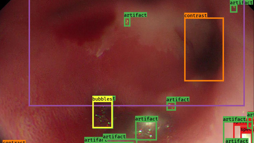
Cancer detection is made even more challenging by artefacts in the endoscopy video such as bubbles, debris, overexposure, light reflection and blurring, which can obscure key features and hinder efforts to automatically analyse endoscopy videos.

In an effort to improve the quality of video endoscopy, a team of researchers from the Institute for Biomedical Engineering (Sharib Ali and Jens Rittscher), the Translational Gastroenterology Unit (Barbara Braden, Adam Bailey and James East) and the Ludwig Institute for Cancer Research (Felix Zhou and Xin Lu) have developed a deep-learning framework for quality assessment of endoscopy videos in near real-time.

This framework, published in the journal Medical Image Analysis, is able to reliably identify six different types of artefacts in the video, generate a quality score for each frame and restore mildly corrupted frames.

Frame restoration can help in building visually coherent 2D or 3D maps for further analysis. In addition, providing quality scores can help trainees to assess and improve their endoscopy screening performance.

Future work aims to employ real-time computer algorithm-aided analysis of endoscopic images and videos, which will enable earlier identification of potentially cancerous changes automatically during endoscopy.

This work was supported by the NIHR Oxford Biomedical Research Centre, the Engineering and Physical Sciences Research Council, the Ludwig Institute for Cancer Research and Health Data Research UK.

← New melanoma drug a step closer to the clinic
IBD treatments do not increase susceptibility to COVID-19, study finds →

Other news

News Categories

News by Month

See all news

Subscribe to the Oxford BRC Newsletter

Keep informed about the work of the Oxford BRC by subscribing to our Mailchimp e-newsletter. It is produced several times a year and delivers news and information about upcoming events straight to your inbox.

Subscribe Now

Oxford BRC on Social Media

  • Facebook
  • LinkedIn
  • Twitter
  • YouTube

Feedback

We’d love to hear your feedback. Please contact us at obrcenquiries@ouh.nhs.uk

  • Data Control and Privacy
  • Accessibility
  • Our Partners
  • Disclaimer
  • Contact

Copyright © 2023 NIHR Oxford Biomedical Research Centre