
International researchers, led by a team at the University of Oxford, have demonstrated how an AI technology can measure inflammation in the heart’s arteries using low-radiation CT heart scans typically used to measure calcium score.
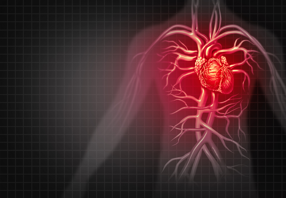
Inflammation can damage the heart by destabilising blood vessel walls, stiffening cardiac muscle and disrupting normal blood flow, so spotting it early could identify more people at risk of a heart attack and allow time for preventive intervention.
The study, which involved researchers from 27 institutions across the UK, USA, Europe and Australia, was published in the Journal of the American Medical Association (JAMA).
The team at the Acute Multidisciplinary Imaging and Interventional Centre (AMIIC) in the Radcliffe Department of Medicine, who are supported by the NIHR Biomedical Research Centre: Oxford and the British Heart Foundation, have already shown that AI technology can detect changes in the fat tissue around the heart’s arteries that are not visible to the human eye. These changes, which can be measured using a Fat Attenuation Index (FAI) Score, are an indicator of inflammation in the arteries.
Until now, it has only been possible to measure inflammation in this way using coronary CT angiography (CCTA), a detailed scan requiring patients to be injected with a special contrast dye to enhance visibility of the vessels in the heart.
Earlier discoveries from this research programme led to the development of CaRi-Heart®, an AI-enabled analysis of CCTA scans that measures coronary inflammation and is available for clinical use through Caristo Diagnostics, a University of Oxford spin-out company.
The new AI model has been trained to extract information about coronary inflammation from non-contrast CT scans already widely used to measure coronary calcium and assess future heart disease risk.
This means that inflammation could potentially be measured from routine scans that have already been obtained in individuals without any cardiovascular symptoms, without having to resort to a procedure involving contrast dye, additional radiation or scanning time.
This could help clinicians identify more people at increased risk of heart attack and intervene earlier with preventive measures, including cholesterol-lowering medicines, blood pressure treatment and lifestyle changes.
Finding hidden cardiovascular risk
In the new study, the team at AMIIC, which is based in the John Radcliffe Hospital, analysed non-contrast CT scans and health records from participants in the international ORFAN study (The Oxford Risk Factors and Non-Invasive Imaging Study). They analysed scans from 19,110 patients, following them and looking at their outcomes for a median of 6.7 years.
During follow-up, 536 patients sadly died from cardiovascular causes and 1,727 experienced a major cardiovascular event. Forty-two per cent of the cardiovascular deaths and 51% of the major cardiovascular events in the study population occurred in people with mild or no coronary calcification.
The team trained the new AI system using data from 6,818 patients and independently tested it in the 19,110-patient study population. The inflammation measurement obtained by the AI tool from non-contrast CT scans predicted cardiovascular risk independently of coronary calcium and performed comparably to the established inflammation measurement obtained using the more detailed contrast-enhanced coronary CT angiography.
The research has also revealed more about the interplay between calcium and inflammation in heart disease. Standard non-contrast CT scans usually measure calcium deposits in the arteries supplying the heart. Higher calcium levels generally indicate more coronary artery disease and greater cardiovascular risk.
The findings in this study were particularly striking in people with a calcium score of zero. Among those who subsequently died from cardiovascular causes, the AI-derived inflammation score identified almost 45% as being at high or very high risk. This shows that someone can have little or no coronary calcium but still have active inflammation and an increased risk of future cardiovascular disease, because inflammation can contribute to the development and rupture of fatty plaques that cause heart attacks.

Dr Kenneth Chan, joint first author of the study and British Heart Foundation clinical research fellow at the University of Oxford, explained: “A calcium score of zero is reassuring, but it does not mean that someone has no risk of a future heart attack.
“Our study shows that inflammation can reveal risk that calcium scoring alone may miss. The important advance is that we can now detect this inflammation from a routine, non-contrast calcium scan. That potentially opens the door to using coronary inflammation in primary prevention, helping us find people at increased risk before they develop more advanced heart disease.”
Next steps
Further research is needed before the technology could be widely used in primary prevention or screening.
Professor Charalambos Antoniades, British Heart Foundation Chair of Cardiovascular Medicine at the University of Oxford and senior author of the study, said: “This is potentially a game-changer for cardiovascular prevention. Calcium tells us about disease that has already developed, while inflammation can reveal disease activity that may otherwise remain hidden.
“Bringing these two pieces of information together could help identify people at risk much earlier and target preventive treatment to those who need it most.”
This study is the latest step in more than a decade of BRC Oxford-supported research into coronary inflammation at the University of Oxford. The translation of scientific discoveries from this research programme into clinical technologies has been taken forward by Caristo Diagnostics.