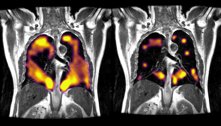
A study by Oxford and Sheffield researchers using a cutting-edge method of imaging has identified persistent damage to the lungs of COVID-19 patients at least three months after they were discharged from hospital, and for some patients even longer.

This damage was not detected by routine CT scans and clinical tests, and the patients would consequently normally be told their lungs are normal.
Further early research by the team has shown that patients who have not been hospitalised with COVID-19 but who are experiencing long-term breathlessness may have similar damage in their lungs, and a larger study is needed to confirm this.
In a paper published in Radiology, the world’s leading radiology journal, the researchers from Oxford and Sheffield said that hyperpolarised xenon MRI (XeMRI) scans had found abnormalities in the lungs of some COVID-19 patients more than three months – and in some cases, nine months – after leaving hospital, when other clinical measurements were normal.
The study’s Principal Investigator Prof Fergus Gleeson, Professor of Radiology at the University of Oxford and Consultant Radiologist at Oxford University Hospitals (OUH) NHS Foundation Trust, said: “Many COVID-19 patients are still experiencing breathlessness several months after being discharged from hospital, despite their CT scans indicating that their lungs are functioning normally.
“Our follow-up scans using hyperpolarised xenon MRI have found that abnormalities not normally visible on regular scans are indeed present, and these abnormalities are preventing oxygen getting into the bloodstream as it should in all parts of the lungs.”
Professor Jim Wild, Head of Imaging and NIHR Research Professor of Magnetic Resonance at the University of Sheffield, said: “The findings of the study are very interesting. The 129Xe MRI is pinpointing the parts of the lung where the physiology of oxygen uptake is impaired due to long standing effects of COVID-19 on the lungs, even though they often look normal on CT scans.
“It is great to see the imaging technology we have developed rolled out in other clinical centres, working with our collaborators in Oxford on such a timely and clinically important study sets a real precedent for multi-centre research and NHS diagnostic scanning with 129Xe MRI in the UK.”
The study, which is supported by the NIHR Oxford Biomedical Research Centre (BRC), has now begun testing patients who were not hospitalised with COVID-19 but who have been attending long COVID clinics.

“Although we are currently only talking about early findings, the XeMRI scans of non-hospitalised patients who are breathless – and 70% of our local patients with Long COVID do experience breathlessness – may have similar abnormalities in their lungs. We need a larger study to identify how common this is and how long it will take to get better.” Prof Gleeson explained.
“We have some way to go before fully comprehending the nature of the lung impairment that follows a COVID-19 infection. But these findings, which are the product of a clinical-academic collaboration between Oxford and Sheffield, are an important step on the path to understanding the biological basis of long COVID and that in turn will help us to develop more effective therapies.”
OUH are in the process of upgrading their MRI scanning capabilities, thanks to support from: the medical‑imaging technology company Polarean Imaging, who are working towards providing a new state-of-the-art polariser; from GE, who provide the team with the Xe-MRI specialty imaging hardware; and from the UK Government’s new equipment fund for imaging capital, with which a more up-to-date magnet has been purchased.
The XeMRI research in Oxford has been co-funded through the National Institute for Health Research (NIHR), University of Oxford COVID Research Funds and Innovate UK, through NCIMI (the National Consortium of Intelligent Medical Imaging). Its CEO, Dr Claire Bloomfield, said: “It’s brilliant to see the collaborative research outputs of the long COVID xenon work published in Radiology. It’s an important study that highlights how intelligent imaging can identify ‘hidden’ impacts for COVID patients, and which is now unlocking wider funding and partner support.
“Our hope is that this work can be further expanded to support more patients living with lung damage post-COVID-19, which had previously gone undetected and support detection both with and without Xenon.”
The study – C-MORE-POST in Oxford and MURCO in Sheffield – forms part of the University of Oxford’s C-MORE (Capturing the MultiORgan Effects of COVID-19) study, which feeds into the major national follow-up study PHOSP-COVID, led by the University of Leicester, which is investigating the long-term effects of COVID-19 on hospitalised patients.