
Oxford researchers have identified a striking immune cell signature that may help to explain what happens in the early stages of idiopathic pulmonary fibrosis (IPF), a serious condition that causes scarring of the lungs and makes breathing difficult.
IPF is usually diagnosed after significant lung scarring has already occurred. Current treatments can slow the disease, but they cannot reverse scarring. These findings offer clues to how the disease develops before extensive lung scarring happens and may help to identify new ways to detect and treat the condition sooner.
The study, published in Cell Reports, was conducted by researchers at the University of Oxford’s MRC Translational Immune Discovery Unit, supported by the National Institute for Health and Care Research (NIHR) Biomedical Research Centre: Oxford and the Medical Research Council.
They used cutting-edge single-cell and spatial technologies to map a major group of immune cells (monocytes and macrophages) across the blood, alveolar surface and lung tissue of patients with IPF. These cells typically help the body respond to infection, but they can also influence inflammation and tissue repair.
The research team found two distinct patterns of gene activity in these immune cells in the disease: one associated with fibrotic remodelling — the process by which lung tissue becomes scarred and its normal structure is disrupted, and the other driven by type I interferon activation, which was found in better-preserved and less fibrotic regions of the lung.
Type I interferons are best known for their role in antiviral immunity, making their prominence in IPF an unexpected finding.
Professor Ling-Pei Ho, the NIHR BRC: Oxford’s Theme Lead for Respiratory Medicine, who led the study, said: “What surprised us was how consistently this interferon signal appeared in the less fibrotic parts of the lung. It was present at the alveolar surface, in relatively preserved regions of lung tissue, and in circulating monocytes from patients with milder disease.”

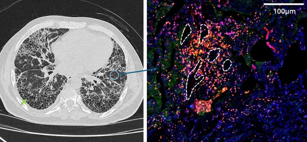
To complement their single-cell RNA sequencing analysis of 108 IPF and control lung samples, Professor Ho’s team used Xenium spatial transcriptomics – a method of genetic profiling that shows cell types and their locations – to generate a high-resolution, unbiased single-cell map of IPF lung tissue.
This revealed reproducible cellular ‘neighbourhoods’ across different patients and showed that interferon-activated alveolar macrophages and monocytes were more plentiful in areas with less fibrotic remodelling.
The same pattern extended beyond the lung. Blood monocytes from people with IPF showed heightened responsiveness to interferon, while higher expression of interferon-related genes was associated with milder disease.
Professor Ho added: “Current treatments target fibrosis once scarring is already established. Our findings raise the possibility of therapeutically targeting immune pathways that are active at earlier stages of fibrosis and in patients with earlier disease before irreversible remodelling of the lungs.”